

Imaging
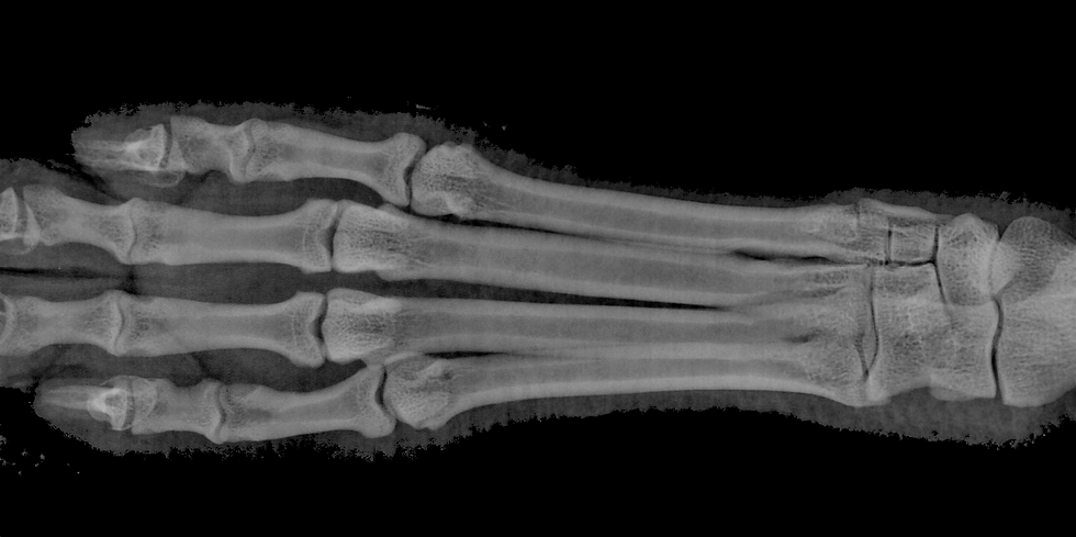
Assessment of cortical bone geometry and cancellous bone architecture is an essential component of our lab. We use several techniques for these assessments including digital radiography, dual energy x-ray absorptiometry, peripheral quantitative computed tomograph, micro-computed tomography.
Major equipment, shared among many labs at IUSM, include:
-
X-ray: Micro Photonics PiXarray 100 digital x-ray machine
-
DEXA: PixiMus II densitomiter (Lunar Corp)
-
pQCT Norland Stratec XCT Research SA+, XCT-2000, and XCT-3000 pQCT machines
-
MicroCT: Skyscan 1172 and 1176 high-resolution desktop micro-computed tomography machines and all associated software


Histology
Assessment of tissue cellular dynamics and damage represent core techniques of most of our studies. We use several techniques for these assessments including:
-
Dynamic histomorphometry to assess mineral apposition rate, minerlaizing surface, and bone formation rate
-
Static histomorphometry to assess osteoblast/osteoid, osteoclast and osteocyte parameters
-
Microdamage assessment on bulk stained tissue
Major equipment in our histology laboratory, that services many labs at IUSM, includes:
-
Microtomes: Reichert-Jung 2050 Supercut; Jung 820 Histocut (2); Reichert-Jung Polycut microtome
-
Sectioning saws: Buehler Isomet Low Speed sectioning saws (2); DDK diamond wire sectioning saws (5)
-
Processors: Shandon automatic processor (PathCenter, Shandon); Embedding Center (Tissue-tek, Sakura); automatic stainer
-
Microscopes:
-
Nikon Optiphot research microscope for brightfield, epifluorescene
-
LEICA research photomicroscope with brightfield
-
-
Image Analysis Systems: Bioquant Osteo image analysis system with CCD camera attachment.
-
The laboratory also has full access to a Reichert-Jung cryostat, several ultramicrotomes, and equipment for electron microscopy.


Mechanics
The ultimate measure of any intervention is how it affects the mechanical properties. We use several techniques for assessing the mechanical properties of bone including:
-
Compression testing of vertebra
-
Reduced platen compression (RPC) of trabecular bone
-
3-point bending of long bones and ribs
-
4-point bending of cortical beams
-
Compression/Shear testing of femoral neck
-
Referenc point indentation (in vivo and ex vivo)
Major equipment in our lab, as well as that housed in the Dept of Biomedical Engineering, includes:
-
Low force (<1000 N) testing machine (Test Resources, 100P225)
-
High force (>1000 N) testing machine (MTS, Alliance RT/5 and Bionix)
-
Reference point indenter (BioDent H, Active Life Technologies)

